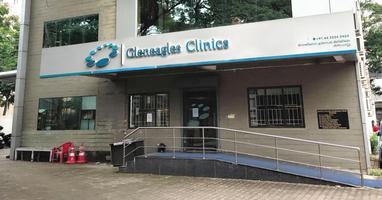
Interventional Radiology at Gleneagles Global Hospitals offers a full range of diagnostic services and therapies for the patients. We take pride in presenting a team of skilled radiologists who use innovative techniques to perform the treatment. Our interventional radiologists are experts in doing interventions in people of all ages. They are highly skilled and are trained from renowned institutions from different parts of the world.
Our physicians, nurse-practitioners, and technicians are trained in special techniques and use advanced tools and equipments for the treatment of the patients. The wards and surgical rooms are well equipped with a good infrastructure focusing on the patients’ safety and comfort. Every test and procedure performed is well organized and the results are accurate; it is the togetherness of the team that brings out great results.
We aim at providing best service and comfort to the patients and help them recover and support them through their medical journey. We not only diagnose but also treat patients using least invasive techniques. The treatments offered at Gleneagles Global Hospitals minimize the patient risk and help them recover soon and lead a healthy life.
Some of the interventional radiology procedures include:
Gleneagles Global Hospitals India is the best Interventional Radiology hospital in India providing advanced care for all types of cancers. The hospital employs Best Interventional Radiologist in India to deliver the finest care and advanced treatments. State-of-the-art infrastructure, diagnostics facilities and the best radiologists in India makes us the best hospital for interventional radiology in India.
Radioembolization is a minimally invasive procedure that is a combination of embolization and radiation therapy to treat liver cancer. It is a technique similar to chemoembolization. It provides localized treatment to the primary or the metastatic liver tumors. Medication filled with tiny glass or resin beads filled with the radioactive isotope yttrium Y-90 are injected into the arteries that lead to the tumors. These beads block the supply of blood to the cancer cells and deliver a high dose of radiation to the tumor without damaging the healthy cells. This enables pin point delivery of radiation to the particular tumor, protecting the surrounding normal tissues.
The doctor begins the procedure by inserting an intravenous line through which sedative medication is given. A catheter is inserted through the skin into the femoral artery, a large groin blood vessel, which is guided to the treatment site. Once the catheter is positioned in the hepatic artery that is feeding the tumor, the tiny glass beads are injected through the catheter to the targeted site. At the end of the procedure, the catheter is removed and the opening of the skin is covered with a dressing.
Tumor Ablation is a treatment involving the use of heating, freezing, or substances such as acetic acid or ethanol which are directed into the tumors to kill the cancer cells. It is relatively a new technique used for the treatment of cancer. A special probe called a radiofrequency electrode is inserted into the tumor with the help of a CT scanner. This is to position the probe in the center of the tumor. This probe destroys the tumor with heat.
During the procedure, using a CT scanner, a small needle-like device is inserted into the tumor through the skin. When the probe is inserted into the cancer, it is attached to a generator which burns or freezes the cancer. This procedure does not damage or destroy the normal healthy cells.
Angiogram is an X-ray test which uses a contrast dye (iodine dye) and camera to diagnose the blockages, narrowing and other blood vessel problems. This test is also referred to as Angiography and the dye that is used is to make the blood vessels visible. It is one of the most common tests performed to show the presence of any heart disease and also the degree of its severity.
During an angiogram, a thin tube called a catheter is placed into a blood vessel in the femoral artery or vein or in the brachial artery or vein. The catheter is guided to be area which is to be studied. An iodine dye is injected into the vessel for a clear visible blood vessel on the X-ray. This method is known as conventional or catheter angiogram. These X-rays can also be stored as pictures in the computer.
Angioplasty is a surgical repair used to remove the fat or cholesterol clogged in the coronary artery of the heart. This procedure is also used to widen the narrowed blood vessels for easily flow of blood.
A thin plastic sheath is inserted into the artery, sometimes through the groin or through the arm and the catheter is passed through the sheath and guided up a blood vessel and then to the arteries surrounding the heart. A small amount of contrast dye is injected into the blood vessel through the catheter. The dye is used to get a clear picture of the blood vessel on the X-ray when the blood moves through the blood vessel.
TACE is a minimally invasive, non-surgical procedure used to treat malignant lesions in the Liver (Liver cancer) that cannot be treated with surgery. The procedure is done with a catheter to deliver both Chemotherapy Medication and embolization materials into the blood vessels that leads to tumor.
It works by blocking the blood supply to tumor. Once blood supply is blocked (Embolized) the Chemotherapy works better on the tumor.
TACE can be helpful for:
Advantages of TACE:
This treatment option is currently performed by few doctors only. We are proud to announce that we have Dr. M C Uthappa – Director, Interventional Radiology and Interventional Oncology, with us. Dr. Uthappa has an experience of over 13500 major interventional radiology cases. He is an expert in all types of interventional radiology procedures for Liver Cancer and these includes TARE, Percutaneous Ethanol Injection(PEI), RFA, TACE, DC bead Embolisation, Portal vein Embolisation and Radio Embolisation (SIRT) .
TARE is a trans-catheter intra-arterial procedure performed by the interventional Radiologist for the treatment of primary and secondary hepatic cancers. In TARE procedure, the radioactive substance is injected through the hepatic artery into the liver tumor directly with the help of a catheter inserted into a peripheral artery. The injected radioactive substance gets deposited in the tumor tissue and then locally emits radiation which causes the destruction of the tumor cells.
TARE can be helpful for:
Benefits of TARE:
This treatment option is currently performed by few doctors only. We are proud to announce that we have Dr. M C Uthappa – Director, Interventional Radiology and Interventional Oncology, with us. Dr. Uthappa has an experience of over 13500 major interventional radiology cases. He is an expert in all types of Interventional radiology procedures for Liver Cancer and these includes TARE, Percutaneous Ethanol Injection(PEI), RFA, TACE, DC bead Embolisation, Portal vein Embolisation and Radio Embolisation (SIRT) .